Over 2,000 years ago Hippocrates, the father of medicine said: “All disease begins in the gut”. Healthy digestion and a healthy microbiome are fundamental to health. The digestive tract is a long tube that goes from the mouth to the anus. It is composed of several organs and accessory organs that work together to intake, break down and absorb food as well as excrete waste material. Each organ of the digestive tract can be affected by dysfunction from gastroesophageal reflux to H. Pylori infections, leaky gut, malabsorption syndrome, maldigestion, food allergies, celiac disease, irritable bowel syndrome, inflammatory bowel disease, etc. It is important to know that digestive issues are not confined to the affected organ, but that they have repercussions for the entire system. Moreover, digestive dysfunction causes maladies that are not exclusively relegated to the organs of digestion. Research shows that several conditions are caused by or correlated with unhealthy microbiome and digestive dysfunction: obesity, type-2 diabetes (Fan & Pedersen, 2021), connective tissue disease (CTD) (Bizzaro et al., 2003), systemic lupus erythematosus (SLE), Grave’s disease (Shor et al., 2012), just to name a few.
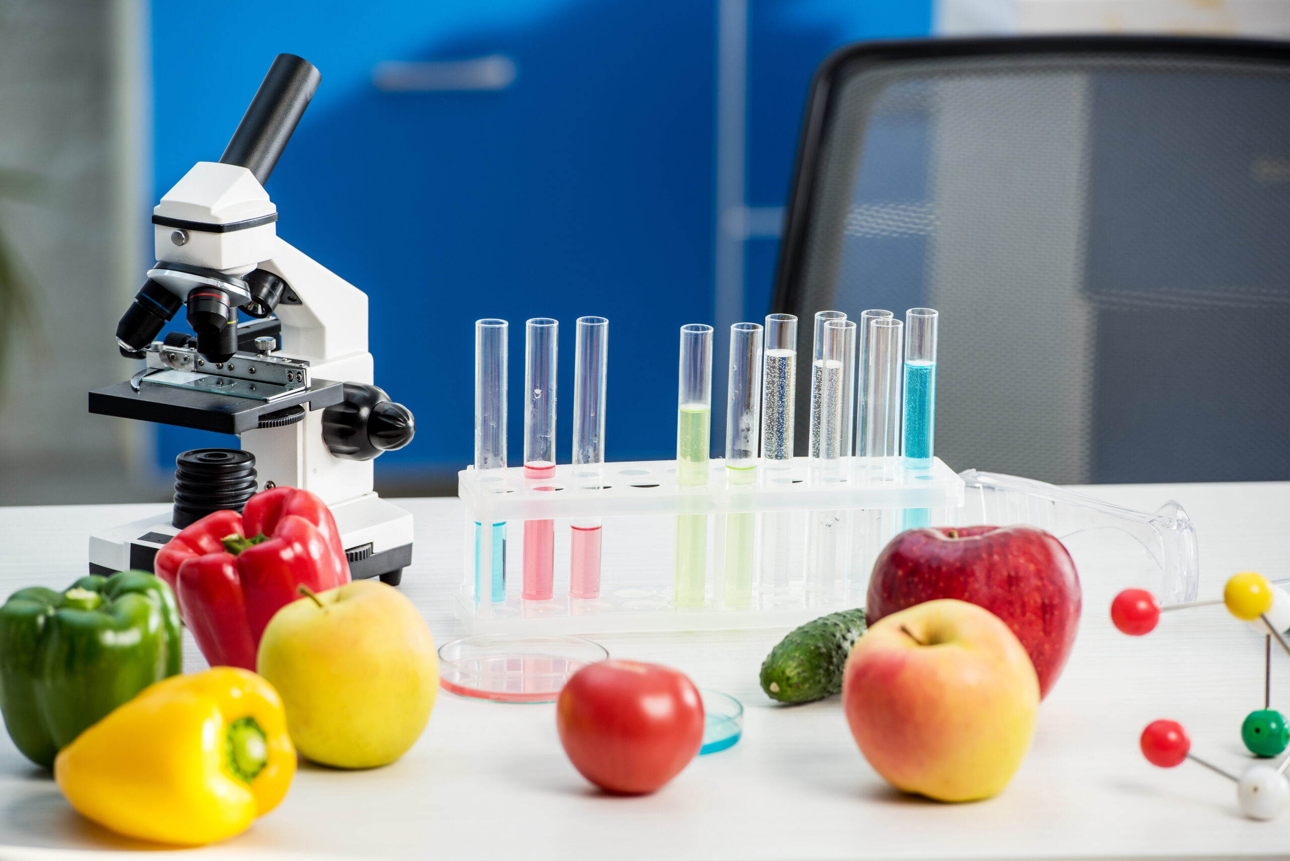
What Laboratory Tests Are Available Today?
There are several laboratory tests available to test gastrointestinal function. Stomach acid can be measured with a Heidelberg capsule (Lord & Bralley, 2012). Pepsin can be tested via a saliva test (Strugala et al., 2015). Fecal and plasma tests can be used to measure pancreatic output of protease and lipase. A fecal fat test can reveal impaired liver or gallbladder function. Stool cultures, DNA stool test, comprehensive stool digestive analysis (CSDA), fecal butyrate testing are tools used to assess colon function (Lord & Bralley, 2012). Colonoscopy, barium enema, magnetic resonance imaging (MRI), computed cosmography scan (CT scan), defecography, ultrasounds and other imaging tests are also available to assess colon health (Digestive Diagnostic Procedures). A hydrogen-methane breath test is used to diagnose small intestinal bacterial overgrowth (SIBO).
Several tests are available to test for food allergies and intolerances: increased levels of IgA can be measured through feces, urine and serum analysis and can reveal the presence of gut inflammation, celiac disease, mucosal infection, food allergies, and other inflammatory conditions (Breedveld & van Egmond, 2019). Serum IgE and IgG levels can be checked to test for food allergies, infections and inflammatory diseases (Mayo Clinic Labs). According to the Genova Diagnostics website, high levels of IgG antibodies can also indicate the presence of leaky gut syndrome.
What Is The Helicobacter Test?
The test I chose for this essay is the Helicobacter Pylori Stool Antigen EIA. The American Gastroenterological Association (AGA) (Talley et al., 2005) and the American College of Gastroenterologists (ACG) (Chey et al., 2007) consider the H. Pylori stool antigen to be superior to the serum testing.
H. Pylori is a bacterium that inhabits the stomach, usually without causing any disease. According to Iisashi et al. (2015) H. Pylori can suppress inflammatory bowel disease (IBD), and it is linked to a reduced incidence of asthma. A study from Talebi Bezmin Abadi (2014) even suggested that an eradication of H. Pylori contributes to an increase of GERD. It is still unknown why, in certain people, H. Pylori colonies wreaks havoc in the stomach, causing stomach ulcers, gastric inflammation, stomach cancer and gastric mucosa-associated lymphoid-tissue lymphoma (Yang it al., 2014). Symptoms associated with H. Pylori infections are burping, bloating, nausea, gastritis presenting with pain and a burning sensation, loss of appetite and weight loss (Mayo clinic, 2017). A patient that presents with these symptoms should be tested for H. Pylori infection. The stool antigen EIA test looks for the present of antigens that reveal the presence of H. Pylori. Certain medications like antibiotics and acid blockers can interfere with the test results; therefore, patients are asked to discontinue the use to these medications for one to two weeks prior to testing.
References
Bizzaro, N., Villalta, D., Tonutti, E., Tampoia, M., Bassetti, D., & Tozzoli, R. (2003). Association of celiac disease with connective tissue diseases and autoimmune diseases of the digestive tract. Autoimmunity reviews, 2(6), 358–363. https://doi.org/10.1016/s1568-9972(03)00055-7
Bravo, D., Hoare, A., Soto, C., Valenzuela, M. A., & Quest, A. F. (2018). Helicobacter pylori in human health and disease: Mechanisms for local gastric and systemic effects. World journal of gastroenterology, 24(28), 3071–3089. https://doi.org/10.3748/wjg.v24.i28.3071
Breedveld, A., & van Egmond, M. (2019). IgA and FcαRI: Pathological Roles and Therapeutic Opportunities. Frontiers in Immunology, 10. https://doi.org/10.3389/fimmu.2019.00553
Chey WD, Wong BC; Practice Parameters Committee of the American College of Gastroenterology. American College of Gastroenterology guideline on the management of Helicobacter pylori infection. Am J Gastroenterol. 2007;102:1808-1825.
Fan, Y., & Pedersen, O. (2021). Gut microbiota in human metabolic health and disease. Nature reviews. Microbiology, 19(1), 55–71. https://doi.org/10.1038/s41579-020-0433-9
Gastrointestinal Test | helicobacter pylori Stool Antigen EIA. (n.d.). Www.gdx.net. Retrieved July 16, 2021, from https://www.gdx.net/product/helicobacter-pylori-stool-antigen-eia-test
IGE – Clinical: Immunoglobulin E (IgE), Serum. (n.d.). Www.mayocliniclabs.com. https://www.mayocliniclabs.com/test-catalog/Clinical+and+Interpretive/8159
Iizasa, H., Ishihara, S., Richardo, T., Kanehiro, Y., & Yoshiyama, H. (2015). Dysbiotic infection in the stomach. World journal of gastroenterology, 21(40), 11450–11457. https://doi.org/10.3748/wjg.v21.i40.11450
Lord, R. and Bralley, J., n.d. Laboratory evaluations for integrative and functional medicine. 2nd ed. (2012), Metametrix institute.
Lu, P. J., Hsu, P. I., Chen, C. H., Hsiao, M., Chang, W. C., Tseng, H. H., Lin, K. H., Chuah, S. K., & Chen, H. C. (2010). Gastric juice acidity in upper gastrointestinal diseases. World journal of gastroenterology, 16(43), 5496–5501. https://doi.org/10.3748/wjg.v16.i43.5496
Mayo Clinic. (2017). Helicobacter pylori (H. pylori) infection – Symptoms and causes. Mayo Clinic; https://www.mayoclinic.org/diseases-conditions/h-pylori/symptoms-causes/syc-20356171
Shor, D. B., Orbach, H., Boaz, M., Altman, A., Anaya, J. M., Bizzaro, N., Tincani, A., Cervera, R., Espinosa, G., Stojanovich, L., Rozman, B., Bombardieri, S., Vita, S. D., Damoiseaux, J., Villalta, D., Tonutti, E., Tozzoli, R., Barzilai, O., Ram, M., Blank, M., … Shoenfeld, Y. (2012). Gastrointestinal-associated autoantibodies in different autoimmune diseases. American journal of clinical and experimental immunology, 1(1), 49–55.
Strugala, V., Woodcock, A. D., Dettmar, P. W., Faruqi, S., & Morice, A. H. (2015). Detection of pepsin in sputum: a rapid and objective measure of airways reflux. European Respiratory Journal, 47(1), 339–341. https://doi.org/10.1183/13993003.00827-2015
Talebi Bezmin Abadi A. (2014). Helicobacter pylori: A Beneficial Gastric Pathogen?. Frontiers in medicine, 1, 26. https://doi.org/10.3389/fmed.2014.00026
Talley NJ; American Gastroenterological Association. American Gastroenterological Association medical position statement: evaluation of dyspepsia. Gastroenterology. 2005;129:1753-1755.
Yang, J. C., Lu, C. W., & Lin, C. J. (2014). Treatment of Helicobacter pylori infection: current status and future concepts. World journal of gastroenterology, 20(18), 5283–5293. https://doi.org/10.3748/wjg.v20.i18.5283
B vitamins are a class of water-soluble vitamins that are essential to maintain health and to carry out a host of metabolic functions. There are 8 different B vitamins that are necessary for several organs and systems to function optimally. B vitamins are utilized for cellular functioning, carbohydrate metabolism and the production of red blood cells. They are also needed for healthy skin and neurotransmitter formation. Symptoms of B vitamin deficiency vary from fatigue to anemia, nervous system dysfunction, compromised immunity and skin issues. While over consumption of B vitamins from food sources is quite rare and generally not worrisome, dangerously high intake of B vitamin through supplementation leads to hypervitaminosis B. This condition comes with side effects that include liver problems, blurry vision, high blood sugar, and numbness.
What Is Folate?
Folate is needed to produce healthy red blood cells; it reduces the risk of neural tube defects such as spina bifida and, together with vitamin B12 and vitamin C, is an important coenzyme in the synthesis of nucleic acids and the metabolism of amino acids. Optimal folate intake helps in the prevention of folate deficiency anemia.
The Importance of B12
Vitamin B12 is an important factor for the formation of red blood cells, and it is essential in preventing megaloblastic anemia. Vitamin B12 is important for DNA synthesis. Cellular metabolism is dependent on this vitamin, which has a role in the metabolism of fatty acids as well as amino acid synthesis. Vitamin B12 is also necessary for the absorption of folate, and it is necessary for conversion of carbohydrates into glucose. Vitamin B12 also plays a role in white blood cell formation, affecting immune system function.
What To Know About Vitamin B 6
Vitamin B6 (Pyridoxine) exists in various coenzyme forms (pyridoxal 5 phosphate (PLP) and Pyridoxamine 5 phosphate (PMP)). It plays a role in over 100 enzyme reactions, and it is involved in the metabolism of protein, carbohydrates, and lipids. Vitamin B6 is essential for brain development and for immune function. It is vital to synthesize neurotransmitters and to ensure adequate levels of homocysteine. It is also essential for gluconeogenesis, glycogenolysis, and hemoglobin formation. Vitamin B6 works synergistically with folate and vitamin B12 to reduce homocysteine levels. High level of homocysteine is a risk factor in heart disease. While the mechanisms aren’t fully understood, it appears that high homocysteine levels can damage arteries, leading to atherosclerosis and blood clots.
What You Need To Know About Choline
Choline is an organic, water-soluble compound similar to B vitamins. It is manufactured in the liver, and it is also found in foods such as liver, muscle meat, fish, eggs, beans, wheat germs, and nuts. Choline is essential in metabolism, and it is needed for cell membrane integrity. It is important for DNA synthesis, cell signaling, fat transport and metabolism. Choline is necessary to make acetylcholine, an important neurotransmitter; therefore, it is crucial for the nervous system. Choline is a source of methyl groups and is needed to produce two major phospholipids (phosphatidylcholine and sphingomyelin) crucial to cell membranes.
What is the RDA level for each of the vitamins? What is the upper limit for each of the vitamins and What are the signs or symptoms of deficiency and toxicity for each of the vitamins?
The RDA for vitamin B6 varies depending on age, sex, and for women RDA changes also during pregnancy and breastfeeding. RDA for babies from birth to six months is 0.1mg; it is 0.3mg for babies from 7 months to 1 year of age. From 1 to 3 years it is 0.5 mg, 4 to 8 years is 0.6 mg while 4 to 8 years 0.6 mg, 9-13 years is 1.0 mg. RDA for males and females between 19 to 50 years is 1.3 mg.RDA of vitamin B6 for males above 51 years is 1.7 mg and for females it is 1.4 mg.
The RDA for vitamin B12 is dependent on age. The RDA for breastfeeding mothers is 2.8 mcg, while for pregnant teens and women it is 2.6 mcg. Teens and adults are recommended to take 2.4 mg per day of vitamin B12. The RDA for children is 1.2 mcg for the age group 4-8 years, and 1.8 mcg for 9 to 13 years. Infants up to 6 months need 0.4 mcg of vitamin B12 per day; the recommendation for infants 7 to 12 months is 0.5 micrograms per day, and children between 1-3 years require 0.9 mcg per day.
The RDA for folate is also dependent on age. Women and men above 19 years should take 400 mcg of dietary folate equivalents (DFE) per day. The RDA for pregnant women is 600 mcg per day, while RDA for lactating women is 500 mcg. The RDA for people who habitually consume alcohol is at 600 mcg.
The Office of Dietary Supplements states that there is insufficient data to establish the RDA for choline; however, adequate intakes (AI) are available. AI is defined as the “recommended average daily nutrient intake level based on approximations of observed mean nutrient intake by a group (or groups) of apparently healthy people that are assumed to be adequate” (Dietary Reference Intakes: applications in dietary assessment). The AI for individuals above 19 years is 550 mg/day for males and 425 mg/day for females. The AI for pregnant women is 450 mg/day, and for lactating women it is 550mg/day. AI is used as a reference levels when there is not enough evidence to develop an RDA.
Choline deficiency can damage the muscles, liver, and it is linked to the development of nonalcoholic fatty liver disease (NAFLD). Most people in the US consume less than the daily requirement of choline; however, thanks to endogenous production of choline by the liver, deficiency in healthy and non pregnant individuals is rare. High doses of choline can cause dizziness and can lower blood pressure. Choline toxicity can also cause also vomiting, increased sweating, salivation, and it can cause fishy body odor.
Vitamin B 12 deficiency causes fatigue, weakness, constipation, loss of appetite, weight loss, and megaloblastic anemia. It also leads to numbness, balance problems, depression, dementia, and confusion. Vitamin B12 toxicity is rare, and it generally manifests with diarrhea, itching, blood clots, numbness of the extremities, and allergic reactions.
Excessive folate intake masks the symptoms of vitamin B 12 deficiency and can cause damage to the nervous system. Its deficiency mainly leads to folate deficiency anemia, diarrhea, gray hair, peptic ulcer, poor growth, glossitis, and ulcers in the mouth.
It is rare to have an isolated deficiency of vitamin B6. While uncommon, vitamin B6 deficiency is associated with microcytic anemia, cheilosis, dermatitis, glossitis, depression, confusion, and a weakened immune system. An excess of the vitamin B6 leads to sensory neuropathy, ataxia, skin lesions, photosensitivity, nausea, and heartburn. These signs and symptoms are dose-specific.
Where are these vitamins found in the diet and what may impede availability and absorption? How does the concept of food poverty impact a client’s ability to obtain these vitamins?
Vitamin B6 is found in a variety of foods, including salmon, beef, liver, pork, potatoes, bananas, and avocado. Other sources include fortified cereals, poultry, pistachio nuts, and non-citrus fruits.
Beef liver is a great source of folate, and boiled spinach comes right behind. Other good sources include broccoli, leafy green vegetables, peas, kidney beans, chickpeas, and fortified cereals. Pregnant women should take folate supplements and consume foods high in folate to prevent neural tube defects.
The best sources of vitamin B12 are liver and clams. Other good sources are meat, fish, milk, cheese, eggs, dairy, and fortified cereals.
Dietary sources of choline include meat, poultry, fish, milk, eggs, gravies, salads, nuts and seeds, and wheat germ.
Several factors can hinder the absorption and availability of vitamins. Various medications can affect the absorption of various vitamins like vitamin B6. These medications include anti-seizure drugs, the bronchodilator theophylline, and antibiotics like cycloserine. The mechanisms for drug-nutrient interactions vary as well. For instance, cycloserine increases the urinary loss of pyridoxine. Availability and absorption of vitamin B12 are also affected by various gastrointestinal conditions like ulcers, inflammatory bowel disease and other digestive disorders, surgical conditions like gastrostomy, and, of course, medications. The medications include chloramphenicol, omeprazole, cimetidine, and metformin. It is worth noting B vitamins are also best absorbed from food sources; however, folate supplementation is needed in pregnancy and some studies show that supplemental folate is better absorbed than dietary folate (85% bioavailability of supplemental compared to 50% from food sources). It is important to identify factors that can affect the vitamins’ availability and to develop proper strategies that will ensure that our clients have optimal intake and absorption.
What Happens When Food Poverty Is A Significant Component In Mal-nutrition?
Food poverty is the inability to access or purchase foods that make up a healthy diet. In other words, food poverty reduces access to the healthy foods that provide an abundance of vitamins and minerals. Food poverty is linked to malnutrition, obesity, vitamin deficiencies, weakened immune system and other diseases, and disordered eating (people living in food insecurity tend to eat even when not hungry, as a guard against food uncertainty). Since food poverty translates to limited access to healthy foods, people affected by food poverty do not meet RDAs and AI of vitamins and minerals.
References:
Stipanuk MH, Caudill MA, editors. Biochemical, physiological, and molecular aspects of human nutrition. 4th ed. St. Louis, Mo: Elsevier; 2019. 959 p.
Bjørndal B, Bruheim I, Lysne V, Ramsvik MS, Ueland PM, Nordrehaug JE, et al. Plasma choline, homocysteine and vitamin status in healthy adults supplemented with krill oil: a pilot study. Scandinavian Journal of Clinical and Laboratory Investigation. 2018 Nov 17;78(7–8):527–32.
Kennedy DO. B vitamins and the brain: mechanisms, dose and efficacy—a review. Nutrients. 2016 Feb;8(2):68.
Office of dietary supplements – choline [Internet]. [cited 2020 Nov 15]. Available from: https://ods.od.nih.gov/factsheets/Choline-HealthProfessional/
Office of dietary supplements – vitamin b6 [Internet]. [cited 2020 Nov 15]. Available from: https://ods.od.nih.gov/factsheets/VitaminB6-HealthProfessional/
Office of dietary supplements – vitamin b12 [Internet]. [cited 2020 Nov 15]. Available from: https://ods.od.nih.gov/factsheets/VitaminB12-HealthProfessional/
Vitamins and minerals – B vitamins and folic acid [Internet]. nhs.uk. 2017 [cited 2020 Nov 15]. Available from: https://www.nhs.uk/conditions/vitamins-and-minerals/vitamin-b/
Sobczyńska-Malefora A, Harrington DJ. Laboratory assessment of folate (Vitamin b9) status. Journal of Clinical Pathology. 2018 Nov 1;71(11):949–56.
Siddiqui F, Salam RA, Lassi ZS, Das JK. The intertwined relationship between malnutrition and poverty. Front Public Health [Internet]. 2020 [cited 2020 Nov 15];8. Available from: https://www.frontiersin.org/articles/10.3389/fpubh.2020.00453/full
Proteins differ greatly in their nutritive value, and there are numerous methods used in nutrition science to establish protein quality and bio-availability. Protein quality refers to a protein’s digestibility as well as its amino acid profile and how well the protein is used by the body to perform specific metabolic functions. One way that protein quality can be evaluated is by categorizing proteins as complete (also referred to as high-quality) or incomplete (also referred to as low-quality). Complete proteins contain all the essential amino acids that the body requires from food whereas incomplete proteins lack one or more of the essential amino acids.
Methods To Evaluate Protein Quality
Different methods are used to evaluate protein quality: biological value (BV) and net protein utilization are two of them. These methods not only look at a food’s protein profile, but they also look at how the body utilizes the protein in a specific food. Biological value is a measure of the proportion of absorbed protein from a food which becomes incorporated into the proteins of the organism’s body. BV uses nitrogen to establish how readily the digested protein can be used in protein synthesis. Proteins are the major source of nitrogen in our diet, and BV assumes protein to be the only source of nitrogen. The difference between the amount of nitrogen ingested and the amount of nitrogen excreted tells us how much nitrogen has been incorporated in an organism’s body. The ratio of nitrogen incorporated into the body over nitrogen absorbed gives the biological value. Unlike other measures of protein usability, BV does not consider how readily the protein can be digested and absorbed.
What Is Net Protein Utilization?
Net protein utilization (NPU) also estimates nitrogen retention. Unlike BV, though, this method estimates nitrogen retention by determining the difference between body nitrogen content of animals fed no protein and those fed a test protein. This value divided by the amount of protein consumed is the NPU, which is defined as the “percentage of the dietary protein retained”. While both NPU and BV estimate retained nitrogen, in the calculation of NPU the denominator is the total protein eaten whereas in the calculation of BV it is the amount absorbed.
Protein and Kidney Health As Used In Nutritional Science
Biological value and net protein utilization are commonly used in nutrition science as a guideline for protein choice in diseased states that need to restrict protein intake. Kidney disease is one of these conditions. People suffering from kidney disease need to restrict protein intake. Eliminating protein altogether is not an option, as protein malnutrition would cause even more harm. Therefore, people suffering from kidney disease need to focus on protein foods of high biological value that the body can metabolize efficiently and that yield very little waste. The diet of a person suffering from kidney disease not in dialysis needs to provide around 0.6 to 0.8g per kg of body weight of proteins. At least 50% of proteins needs to be of high biological value (eggs, meat and poultry, fish, and dairy). People on dialysis have higher protein needs that those who are not on dialysis; therefore, patients on peritoneal dialysis need 1.3g per kg of body weight; patients in hemodialysis are recommended 1.2g per kg of body weight.
Sarwar G, Blair R, Friedman M, Gumbmann MR, Hackler LR, Pellett PL, et al. Inter- and Intra-laboratory Variability in Rat Growth Assays for Estimating Protein Quality of Foods. Journal of AOAC INTERNATIONAL. 1984 Sep 1;67(5):976–81
Stipanuk MH, Caudill MA, editors. Biochemical, physiological, and molecular aspects of human nutrition. 4th ed. St. Louis, Mo: Elsevier; 2019. 959 p.
The digestive system breaks down the food we ingest and turns it into energy and waste products. It is composed of the digestive tract and accessory organs. The digestive tract is a hollow tube that goes from mouth to anus, with openings at each end. The accessory organs are the salivary glands, the pancreas, the liver and gallbladder.
Digestion begins in the brain, with the cephalic phase of digestion triggering the release of gastric secretion as a result of the sight, smell and thought of food. Food enters the mouth where the teeth begin the physical break down food. Three sets of salivary glands secrete saliva to moisten the food and help with swallowing.These glands also secrete the enzyme salivary amylase, also known as ptyalin, which begins the chemical breakdown of starch into maltose. Salivary amylase is in contact with food for a very short time, but deficiency of this enzyme creates a burden further down in the small intestine where pancreatic alpha-amylase has to work harder to break down starches.
The Role That The Mouth and Tongue Play In Digestion
The tongue contains taste buds, and it also pushes food toward the back to the mouth for swallowing. When we swallow, the bolus enters the esophagus for passage to the stomach through the cardiac sphincter. The stomach continues the mechanical and chemical breakdown of food. Gastric juice is secreted by millions of gastric glands located in the mucosal lining of the stomach. It is composed of mucus, pepsinogen (the inactive form of the enzyme pepsin), and hydrochloric acid (HCl). HCl activates pepsin from pepsinogen, and it triggers the hormonegastrin to be released into the bloodstream. This hormone signals the brain to produce more gastric juice. HCl and pepsin break down protein into peptides. Pepsin deficiency is generally associated with low levels of HCl, a condition known as hypochloridria. Symptoms include GERD, burping, bloating, gas, nausea and diarrhea, H. Pylori infections, maldigestion, neurological issues, iron-deficiency anemia, vitamin B12 deficiency, calcium, magnesium and protein deficiency.
The Stomach, pH Levels and the Role They Play In Healthy Digestion
The stomach is all about acid (pH of 1.5 – 3.0). HCl is excreted into the stomach at a pH of 0.8. The acid bathes the stomach, disinfects it, kills bacteria and parasites, and activates pepsin for protein digestion. After the stomach churns the bolus and mixes it with gastric juice, the bolus breaks down even more into a paste called chyme, which is released into the upper part of the small intestine (duodenum) through the pyloric sphincter. When chyme enters the duodenum, the acidic pH of the chyme triggers the goblet cells of the small intestine to secrete mucous.
The Small Intestines And Digestion
The small intestine has the dual roles of digestive organ and gland. The intestinal walls secrete two hormones into the bloodstream: secretin and cholecystokinin (CCK). Secretin stimulates the pancreas to release bicarbonate and pancreatic juice, and CCK stimulates the gallbladder to release bile necessary to emulsify and absorb fats. As part of the pancreatic juice, the pancreas first releases sodium bicarbonate to help raise the pH of the chyme to neutral. Once the chyme pH reaches neutral, the enzyme portion of the pancreatic juice is released to complete the chemical digestion of carbohydrates, proteins, and fats. Three major groups of pancreatic enzymes are proteases, pancreatic lipase, and pancreatic alpha-amylase. Other pancreatic enzymes include gelatinase, elastase, deoxyribonuclease, and ribonuclease.
While digestion of protein begins in the stomach, the bulk of this process takes place in the small intestine and is carried out by pancreatic proteases: mainly trypsin and chymotrypsin. These enzymes are stored in the pancreas and released in inactive form (trypsinogen and chymotrypsinogen). When they reach in the lumen of the small intestine, the enzyme enterokinase, present in the intestinal mucosa, converts trypsinogen into its active form, trypsin. When activated, trypsin activates chymotrypsin. Both enzymes break down peptides into tripeptides and dipeptides. The final digestion of tri- and dipeptides into single amino acids is carried out by carboxypeptidase and elastase. Proteases deficiency causes protein maldigestion, protein deficiency, hypoglycemia, weakness, constipation, hair loss, gingivitis, calcium deficiency, tooth decay, and mood swings.
The Pancreas and Digestion
Pancreatic lipase breaks down triglycerides into monoglycerides and free fatty acids. Bile salts are also necessary for lipase to work as well as for the absorption of fatty acids and monoglycerides. Pancreatic lipase deficiency causes fat maldigestion, steatorrhea, abdominal pain, weight loss, fat-soluble vitamin deficiencies, and calcium deficiency.
Pancreatic alpha-amylase breaks down starch into maltose, maltotrios and alpha-limit dextrins. Alpha-amylase deficiency causes carbohydrate maldigestion, malabsorption, gas and diarrhea, and bacterial overgrowth.
By the time chyme leaves the duodenum, it is almost fully digested. Peristalsis moves the absorbable molecules into the jejunum, where millions of villi and microvilli absorb them into the bloodstream, which carries them to the entire body. The leftover chyme from the small intestine (indigestible fibers, bile, water, and sloughed off cells) passes into the large intestine through the ileocecal valve. The large intestine recycles water and the waste material that nourishes the colon cells; it captures any nutrients that are still available, with the help of the bowel flora, and converts the nutrients to Vitamins K/B1/B2/B12 and butyric acid; it forms and expels feces.
References:
des Gachons CP, Breslin PAS. Salivary Amylase: Digestion and Metabolic Syndrome. Curr Diab Rep. 2016 Oct;16(10):102.
This website collects cookies. Please read our Privacy Policy to review the updates about which cookies we use and what information we collect on our site. By continuing to use this site, you are agreeing to our updated privacy policy.