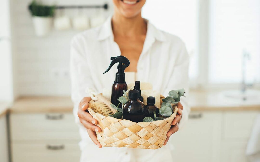
Commercial cleaning products are laden with harsh chemicals that are hazardous to our health and the environment (EWG, 2023). This awareness has led to a growing demand for alternative, eco-friendly solutions. Cleaning your home doesn’t have to involve exposing yourself and your loved ones to harmful substances. There are eco-friendly alternatives on the market, and you can also make cleaning supplies with a few ingredients. I have been using essential oils (EOs) to clean and disinfect my home for a few years, and I am thrilled to share my recipes with you.
EOs are very effective for cleaning. Research shows certain essential oils have antiseptic, antibacterial, and antiviral properties. My favorite essential oils for non-toxic household cleaners are clove, tea tree, lemon, and eucalyptus, but endless possibilities exist. I choose these oils because of the research studies behind them. For example, clove has powerful antiseptic properties. It has been shown to have antibacterial, antiviral, and antifungal effects, making it an effective disinfectant for the home (Bai et al., 2023). A study published in the journal Pathogens found that clove oil has antibacterial activity against multidrug-resistant Streptococcus suis (Wongsawan et al., 2019). Tea tree essential oil is another favorite because of its powerful antiseptic properties. It is effective against many bacteria and viruses, including methicillin-resistant Staphylococcus aureus (MRSA) and influenza (Carson et al., 2002). A study published in the Journal of Hospital Infection found that tea tree oil effectively killed several strains of bacteria commonly found in hospitals, including Staphylococcus aureus and Pseudomonas aeruginosa (Carson et al., 2006). Lemon essential oil is a natural disinfectant with a fantastic, fresh, citrusy scent. It is an excellent disinfectant and has antibacterial and antiviral properties. In 2020, a study by Kumar et al. demonstrated that lemon and geranium essential oils effectively against the SARS-CoV-2/COVID-19 virus.
Last but not least, eucalyptus essential oil has been shown to have antibacterial, antiviral, and antifungal properties. It has been used traditionally as a natural remedy for respiratory infections and is effective against several strains of bacteria and viruses. A systematic review of 113 articles by Elangovan and Mudgil (2023) demonstrated that eucalyptus oil exhibited strong antibacterial activity against several strains of bacteria, including MRSA. I combine these four essential oils to make a pleasant disinfectant spray that is an effective natural alternative to harsh chemical disinfectants.
All-Purpose Cleaner
DIY Recipe:
Ingredients:
8 oz dark glass spray bottle
3/4 cup distilled water
two tablespoons of vinegar
10 drops clove essential oil
10 drops tea tree essential oil
10 drops lemon essential oil
10 drops eucalyptus essential oil
Instructions: Add each ingredient to the bottle and shake vigorously to combine, and my EO disinfectant spray is created!
Floor Cleaner:
DIY Recipe:
Ingredients:
1 cup distilled white vinegar
1 cup water
1 tablespoon castile soap (unscented or a mild scent)
10 drops tea tree essential oil
10 drops lemon essential oil
5 drops eucalyptus essential oil
5 drops clove essential oil
Spray bottle
Instructions:
In a mixing bowl, combine 1 cup of distilled white vinegar and 1 cup of water. Mix in 1 tablespoon of castile soap. Add the essential oils and stir the mixture well. Transfer the mixture to a clean spray bottle.
Notes:
Shake the bottle before each use. Spray the floor lightly and mop as usual. For hardwood floors, spray a small amount directly onto a mop or cloth rather than the floor itself.
Dish Soap:
DIY Recipe:
Ingredients:
1 cup liquid castile soap (unscented or a mild scent)
1 tablespoon washing soda
1 tablespoon vegetable glycerin
10 drops lemon essential oil
5 drops tea tree essential oil
5 drops eucalyptus essential oil
1 cup distilled water
Clean, empty dish soap bottle
Instructions:
In a mixing bowl, pour 1 cup of liquid castile soap. Mix in 1 tablespoon of washing soda and 1 tablespoon of vegetable glycerin. Glycerin helps create a smooth texture and adds moisturizing properties. Add the essential oils and stir well to ensure all ingredients are well combined. Gradually add 1 cup of distilled water while continuing to stir. Pour the mixture into a clean and empty dish soap dispenser or bottle.
Notes:
Shake well before each use. If you have hard water, you may need to add more washing soda to enhance the soap’s effectiveness.
Laundry Detergent:
DIY Recipe:
Ingredients:
1 bar of unscented castile soap
1 cup washing soda
1 cup borax
15 drops tea tree essential oil
15 drops lemon essential oil
10 drops eucalyptus essential oil
5 drops clove essential oil
Glass jar
Instructions:
Grate the bar of castile soap using a cheese grater. You can also cut it into small pieces for easier processing. Combine the grated castile soap, washing soda, and borax in a large mixing bowl. Mix well until all ingredients are evenly distributed. Add the essential oils and transfer the mixture into an airtight container for storage. A glass jar with a lid works well. Use 1-2 tablespoons of the homemade detergent per load, depending on the size of your laundry and the soil level.
Note: Always test DIY solutions in a small, inconspicuous area before use.
References
Bai, J., Li, J., Chen, Z., Bai, X., Yang, Z., Wang, Z., & Yang, Y. (2023). Antibacterial activity and mechanism of clove essential oil against foodborne pathogens. Lebensmittel-Wissenschaft & Technologie, 173, 114249. https://doi.org/10.1016/j.lwt.2022.114249
Carson, C. F., Mee, B. J., & Riley, T. V. (2002). Mechanism of action of Melaleuca alternifolia (tea tree) oil on Staphylococcus aureus determined by time-kill, lysis, leakage, and salt tolerance assays and electron microscopy. Antimicrobial agents and chemotherapy, 46(6), 1914–1920. https://doi.org/10.1128/AAC.46.6.1914-1920.2002
Elangovan, S., & Mudgil, P. (2023). Antibacterial Properties of Eucalyptus globulus Essential Oil against MRSA: A Systematic Review. Antibiotics (Basel, Switzerland), 12(3), 474. https://doi.org/10.3390/antibiotics12030474
Kumar, K. J. S., Vani, M. G., Wang, C., Chen, C., Chen, Y., Lu, L., Huang, C., Lai, C., & Wang, S. (2020). Geranium and Lemon Essential Oils and Their Active Compounds Downregulate Angiotensin-Converting Enzyme 2 (ACE2), a SARS-CoV-2 Spike Receptor-Binding Domain, in Epithelial Cells. Plants, 9(6), 770. https://doi.org/10.3390/plants9060770
Wongsawan, K., Chaisri, W., Tangtrongsup, S., & Mektrirat, R. (2019). Bactericidal Effect of Clove Oil against Multidrug-Resistant Streptococcus suis Isolated from Human Patients and Slaughtered Pigs. Pathogens (Basel, Switzerland), 9(1), 14. https://doi.org/10.3390/pathogens9010014
Consumernotice.org provides comprehensive coverage of the ongoing legal battles surrounding Roundup, a popular herbicide. It outlines the numerous lawsuits filed against Monsanto, now owned by Bayer, by individuals who allege that exposure to Roundup caused them to develop non-Hodgkin lymphoma and other cancers. The article details the significant legal developments, including trial outcomes, settlement offers, and scientific studies regarding the potential carcinogenic effects of glyphosate, the active ingredient in Roundup.
To gain a deeper understanding of these legal proceedings and their implications, read the full article here.
Consumernotice.org offers an in-depth analysis of Roundup, a widely-used herbicide containing glyphosate. It discusses its applications, potential risks, and controversies, particularly concerning its links to cancer. The piece delves into various aspects of glyphosate use in agriculture, its availability in the market, and the ongoing debate in the scientific community about its safety. The article also highlights legal actions and settlements related to Roundup and presents alternatives to using glyphosate-based products.
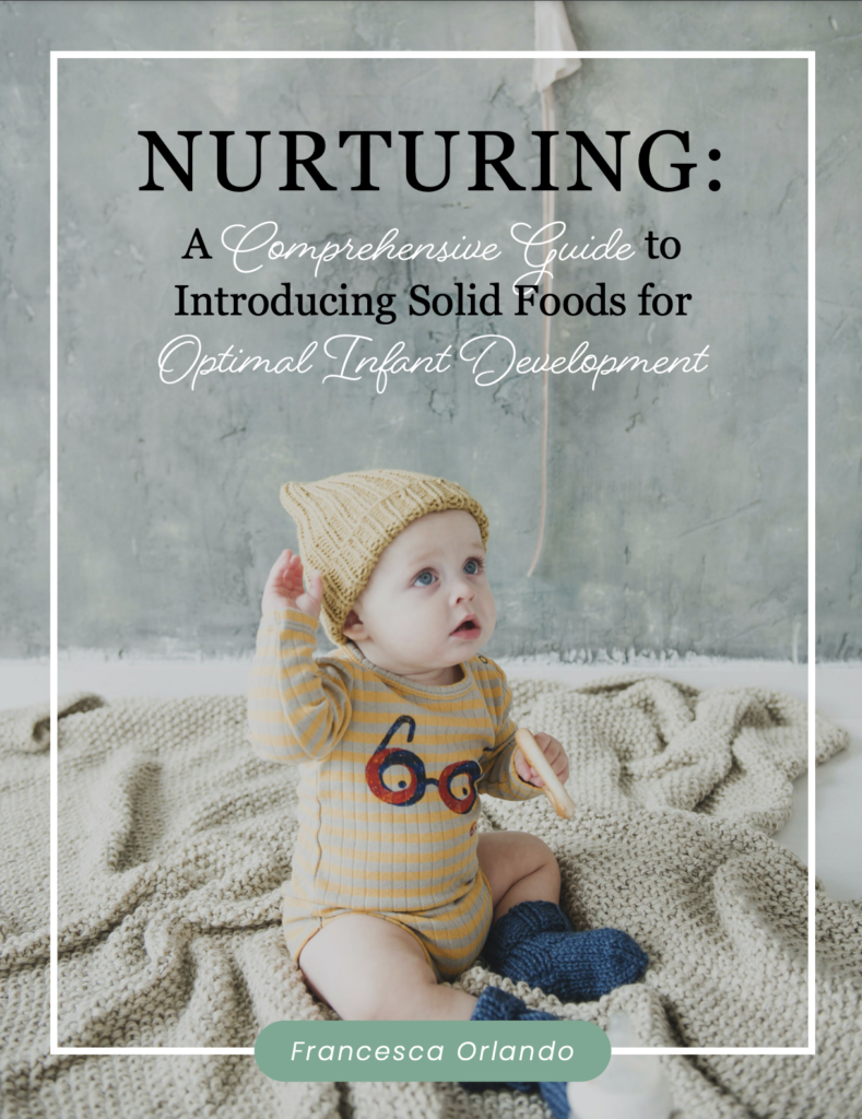
Introducing solid foods to your baby is a momentous occasion, marking a crucial stage in their development. Baby-led weaning (BLW) is a popular approach that encourages self-feeding, allowing infants to explore and enjoy a variety of tastes and textures from an early age.
Baby-led weaning, a term coined by Gill Rapley in 2005, is gaining popularity as a method of introducing solid foods to infants. Rapley, a midwife in the UK, defines baby-led weaning as allowing infants to self-feed appropriately sized whole pieces of food from around six months of age. This approach puts babies in control of what, how much, and how quickly they eat, emphasizing graspable foods they can pick up and hold.
What Are the Benefits of Baby-Led Weaning?
The benefits of baby-led weaning (BLW) are manifold. BLW exposes babies to a wider variety of flavors and textures earlier than traditional purée-fed babies, promoting the development of essential oral motor skills. When done correctly, BLW allows babies to control their food intake, reducing the risk of overeating or undereating. Mealtime experiences become pleasant, as babies can actively participate in the feeding process.
Practical Tips for Successful Baby-Led Weaning:
Set Up a Successful Feeding Environment:
Sit and eat with your baby during mealtimes to serve as a role model.
Ensure safe seating, with babies sitting completely upright for effective feeding.
Let Baby Lead the Way:
Be responsive to baby’s cues and encourage self-feeding.
Embrace the mess and allow babies to explore food using all their senses.
Offer Appropriate Foods:
Use the squish test to determine if a food is safe for the baby.
Advance babies in textures to expose them to a variety of flavors and shapes.
Learn the Difference Between Gagging and Choking:
Recognize the signs of choking and differentiate them from normal gagging.
Get comfortable with gagging as a natural part of the learning process.
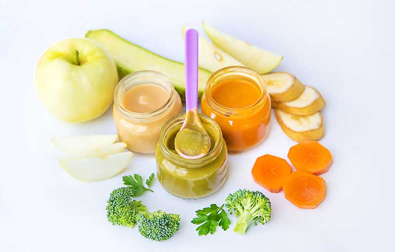
Here are three easy baby-led weaning recipes that are nutritious and suitable for introducing solid foods to your baby:
Avocado and Banana Mash:
Ingredients:
1 ripe avocado
1 ripe banana
Instructions:
Peel and pit the avocado.
Mash the avocado and banana together until you achieve a smooth consistency.
Serve in small, baby-friendly portions.
This recipe introduces healthy fats from avocado and natural sweetness from banana, providing a good mix of nutrients.
Sweet Potato Fingers:
Ingredients:
1 sweet potato
Instructions:
Preheat the oven to 375°F (190°C).
Peel the sweet potato and cut it into finger-sized sticks.
Place the sweet potato sticks on a baking sheet.
Bake for 15-20 minutes or until the sweet potato is tender.
Allow it to cool before serving.
Sweet potatoes are rich in vitamins and minerals, and the finger shape makes it easy for your baby to grasp.
Oatmeal Pancakes:
Ingredients:
1/2 cup rolled oats
1/2 ripe banana, mashed
1/2 cup milk (breast milk, formula, or cow’s milk)
1 egg
Instructions:
In a blender, combine oats, mashed banana, milk, and egg. Blend until smooth.
Heat a non-stick pan over medium heat.
Pour small amounts of batter onto the pan to form mini pancakes.
Cook for 1-2 minutes on each side or until golden brown.
Cool and cut into bite-sized pieces.
These oatmeal pancakes offer a soft texture and are a great way to introduce oats and bananas to your baby.
Remember to always adapt recipes based on your baby’s age and chewing abilities. Supervising your baby while eating is essential, as is ensuring the food is cut into appropriate sizes to prevent choking hazards. If introducing allergenic foods, follow your pediatrician’s advice and introduce one new ingredient at a time, waiting a few days before introducing another.
Safety Measures and Best Practices:
Supervision is Key: Always supervise your baby during meals. This ensures their safety and allows you to observe their eating habits and preferences.
Minimize Choking Hazards: Cut foods into manageable pieces to minimize choking hazards. Avoid small, hard, or round foods that could pose a risk.
Encourage Proper Chewing: Help your baby develop proper chewing skills by offering age-appropriate textures. This can reduce the risk of choking and promote healthy eating habits.
By following these tips and embracing the baby-led feeding approach, parents can make the introduction of solid foods a positive and enjoyable experience for themselves and their little ones. Remember, the goal is to foster a lifelong love for diverse and nutritious foods while ensuring a safe and developmentally appropriate feeding journey.
FAQs Related to Baby-Led Weaning
When can my baby start baby-led weaning?
Your baby should be at least 6 months old.
The tongue thrust instinct should be lost (usually around 6 months).
The baby should sit up independently for 60 seconds and show eagerness to eat.
Follow your baby’s developmental readiness, as recommended by the American Academy of Pediatrics. Breastmilk or formula is sufficient until around 6 months.
Does baby-led weaning increase choking risk?
No, according to the BLISS study.
Introducing various textures through baby-led weaning helps babies become skilled eaters, reducing the likelihood of gagging or choking.
How long does baby-led weaning take?
On average, it may take 2-3 weeks to notice solid food in your baby’s stool.
Begin with one meal per day around 6 months, progress to three meals by 9 months, and reach three meals plus snacks by one year.
What if my baby spits food or doesn’t want to eat it?
Persistence is key. If your baby spits out or pushes away food, continue offering it.
Preferences take time to develop, and it may require several attempts before your baby decides on likes and dislikes.
How do I know if my baby has eaten enough?
Your baby will lose interest in eating when satisfied.
D’Auria, E., Bergamini, M., Staiano, A., Banderali, G., Pendezza, E., Penagini, F., … & Peroni, D. G. Baby-led weaning: what a systematic review of the literature adds on. Italian journal of pediatrics, 44(1), 1-11, 2018.
Introducing the InspiHER’d podcast episode featuring a captivating conversation with Francesca, bringing a fresh perspective on nutrition, philosophy, and cultural differences. In this episode, we dive into the world of holistic nutrition with a passionate expert hailing from Italy, discussing the profound disparities between food in my home country and the United States. Get ready to embark on a journey of discovery as we explore the raw, unfiltered truth behind nutrition and delve into the my mission to promote a healthier and more conscious approach to eating. This episode promises to be an enlightening and thought-provoking experience, offering valuable insights into the power of real, wholesome food and the impact it can have on our lives.
Thanksgiving is a time to gather with loved ones, express gratitude, and enjoy delicious food. While it’s easy to get carried away with indulging in our favorite dishes, it’s important to approach the holiday with mindfulness and gratitude. In this blog post, we will explore the concept of being mindful and grateful at Thanksgiving and provide you with tips to stay happy and healthy while still savoring your favorite foods.
Embrace Mindful Eating: Thanksgiving is the perfect opportunity to practice mindful eating. Slow down, savor each bite, and truly appreciate the flavors, textures, and aromas of the food. By paying attention to your body’s hunger and fullness cues, you can avoid overeating and enjoy your meal more fully.
Prioritize Portion Control: While it’s tempting to load up your plate with everything on the table, practicing portion control is key to maintaining a healthy balance. Start by filling half of your plate with vegetables, a quarter with lean protein, and the remaining quarter with your favorite indulgent dishes. This way, you can enjoy a little bit of everything without going overboard.
Make Healthier Swaps: Thanksgiving dishes can often be heavy on calories and unhealthy fats. Consider making some healthier swaps to lighten up your meal without sacrificing taste. For example, opt for roasted sweet potatoes instead of candied yams, whole grain bread stuffing instead of white bread, and homemade cranberry sauce instead of the canned version loaded with added sugars.
Stay Active: Thanksgiving doesn’t have to be all about food. Incorporating physical activity into your day can help balance out the indulgence. Take a family walk after the meal, engage in a friendly game of touch football, or simply dance to some festive tunes. Not only will this help burn off some calories, but it will also boost your mood and energy levels.
Practice Gratitude: Thanksgiving is a time to express gratitude for the blessings in our lives. Take a moment to reflect on what you’re thankful for and share it with your loved ones. Cultivating an attitude of gratitude can help shift your focus from food to the meaningful connections and experiences that make the holiday special.
This Thanksgiving, let’s approach the holiday with mindfulness and gratitude. By practicing mindful eating, prioritizing portion control, making healthier swaps, staying active, and expressing gratitude, you can enjoy a happy and healthy Thanksgiving while still indulging in your favorite foods. Remember, it’s all about finding a balance that nourishes both your body and soul.
As an integrative nutritionist, I am also grateful for the incredible support of my clients and followers. Your trust and dedication to living a healthful life inspire me every day. I am thankful for the opportunity to be a part of your wellness journey and to provide guidance on your path to optimal health. Your commitment to self-care and your willingness to embrace new ideas and habits is truly commendable. Thank you for allowing me to be a part of your lives.
May this Thanksgiving be a time of joy, gratitude, and connection. Wishing you and your loved ones a healthy and happy holiday season!
This website collects cookies. Please read our Privacy Policy to review the updates about which cookies we use and what information we collect on our site. By continuing to use this site, you are agreeing to our updated privacy policy.