Over 2,000 years ago Hippocrates, the father of medicine said: “All disease begins in the gut”. Healthy digestion and a healthy microbiome are fundamental to health. The digestive tract is a long tube that goes from the mouth to the anus. It is composed of several organs and accessory organs that work together to intake, break down and absorb food as well as excrete waste material. Each organ of the digestive tract can be affected by dysfunction from gastroesophageal reflux to H. Pylori infections, leaky gut, malabsorption syndrome, maldigestion, food allergies, celiac disease, irritable bowel syndrome, inflammatory bowel disease, etc. It is important to know that digestive issues are not confined to the affected organ, but that they have repercussions for the entire system. Moreover, digestive dysfunction causes maladies that are not exclusively relegated to the organs of digestion. Research shows that several conditions are caused by or correlated with unhealthy microbiome and digestive dysfunction: obesity, type-2 diabetes (Fan & Pedersen, 2021), connective tissue disease (CTD) (Bizzaro et al., 2003), systemic lupus erythematosus (SLE), Grave’s disease (Shor et al., 2012), just to name a few.
What Laboratory Tests Are Available Today?
There are several laboratory tests available to test gastrointestinal function. Stomach acid can be measured with a Heidelberg capsule (Lord & Bralley, 2012). Pepsin can be tested via a saliva test (Strugala et al., 2015). Fecal and plasma tests can be used to measure pancreatic output of protease and lipase. A fecal fat test can reveal impaired liver or gallbladder function. Stool cultures, DNA stool test, comprehensive stool digestive analysis (CSDA), fecal butyrate testing are tools used to assess colon function (Lord & Bralley, 2012). Colonoscopy, barium enema, magnetic resonance imaging (MRI), computed cosmography scan (CT scan), defecography, ultrasounds and other imaging tests are also available to assess colon health (Digestive Diagnostic Procedures). A hydrogen-methane breath test is used to diagnose small intestinal bacterial overgrowth (SIBO).
Several tests are available to test for food allergies and intolerances: increased levels of IgA can be measured through feces, urine and serum analysis and can reveal the presence of gut inflammation, celiac disease, mucosal infection, food allergies, and other inflammatory conditions (Breedveld & van Egmond, 2019). Serum IgE and IgG levels can be checked to test for food allergies, infections and inflammatory diseases (Mayo Clinic Labs). According to the Genova Diagnostics website, high levels of IgG antibodies can also indicate the presence of leaky gut syndrome.
What Is The Helicobacter Test?
The test I chose for this essay is the Helicobacter Pylori Stool Antigen EIA. The American Gastroenterological Association (AGA) (Talley et al., 2005) and the American College of Gastroenterologists (ACG) (Chey et al., 2007) consider the H. Pylori stool antigen to be superior to the serum testing.
H. Pylori is a bacterium that inhabits the stomach, usually without causing any disease. According to Iisashi et al. (2015) H. Pylori can suppress inflammatory bowel disease (IBD), and it is linked to a reduced incidence of asthma. A study from Talebi Bezmin Abadi (2014) even suggested that an eradication of H. Pylori contributes to an increase of GERD. It is still unknown why, in certain people, H. Pylori colonies wreaks havoc in the stomach, causing stomach ulcers, gastric inflammation, stomach cancer and gastric mucosa-associated lymphoid-tissue lymphoma (Yang it al., 2014). Symptoms associated with H. Pylori infections are burping, bloating, nausea, gastritis presenting with pain and a burning sensation, loss of appetite and weight loss (Mayo clinic, 2017). A patient that presents with these symptoms should be tested for H. Pylori infection. The stool antigen EIA test looks for the present of antigens that reveal the presence of H. Pylori. Certain medications like antibiotics and acid blockers can interfere with the test results; therefore, patients are asked to discontinue the use to these medications for one to two weeks prior to testing.
References
Bizzaro, N., Villalta, D., Tonutti, E., Tampoia, M., Bassetti, D., & Tozzoli, R. (2003). Association of celiac disease with connective tissue diseases and autoimmune diseases of the digestive tract. Autoimmunity reviews, 2(6), 358–363. https://doi.org/10.1016/s1568-9972(03)00055-7
Bravo, D., Hoare, A., Soto, C., Valenzuela, M. A., & Quest, A. F. (2018). Helicobacter pylori in human health and disease: Mechanisms for local gastric and systemic effects. World journal of gastroenterology, 24(28), 3071–3089. https://doi.org/10.3748/wjg.v24.i28.3071
Breedveld, A., & van Egmond, M. (2019). IgA and FcαRI: Pathological Roles and Therapeutic Opportunities. Frontiers in Immunology, 10. https://doi.org/10.3389/fimmu.2019.00553
Chey WD, Wong BC; Practice Parameters Committee of the American College of Gastroenterology. American College of Gastroenterology guideline on the management of Helicobacter pylori infection. Am J Gastroenterol. 2007;102:1808-1825.
Fan, Y., & Pedersen, O. (2021). Gut microbiota in human metabolic health and disease. Nature reviews. Microbiology, 19(1), 55–71. https://doi.org/10.1038/s41579-020-0433-9
Gastrointestinal Test | helicobacter pylori Stool Antigen EIA. (n.d.). Www.gdx.net. Retrieved July 16, 2021, from https://www.gdx.net/product/helicobacter-pylori-stool-antigen-eia-test
IGE – Clinical: Immunoglobulin E (IgE), Serum. (n.d.). Www.mayocliniclabs.com. https://www.mayocliniclabs.com/test-catalog/Clinical+and+Interpretive/8159
Iizasa, H., Ishihara, S., Richardo, T., Kanehiro, Y., & Yoshiyama, H. (2015). Dysbiotic infection in the stomach. World journal of gastroenterology, 21(40), 11450–11457. https://doi.org/10.3748/wjg.v21.i40.11450
Lord, R. and Bralley, J., n.d. Laboratory evaluations for integrative and functional medicine. 2nd ed. (2012), Metametrix institute.
Lu, P. J., Hsu, P. I., Chen, C. H., Hsiao, M., Chang, W. C., Tseng, H. H., Lin, K. H., Chuah, S. K., & Chen, H. C. (2010). Gastric juice acidity in upper gastrointestinal diseases. World journal of gastroenterology, 16(43), 5496–5501. https://doi.org/10.3748/wjg.v16.i43.5496
Mayo Clinic. (2017). Helicobacter pylori (H. pylori) infection – Symptoms and causes. Mayo Clinic; https://www.mayoclinic.org/diseases-conditions/h-pylori/symptoms-causes/syc-20356171
Shor, D. B., Orbach, H., Boaz, M., Altman, A., Anaya, J. M., Bizzaro, N., Tincani, A., Cervera, R., Espinosa, G., Stojanovich, L., Rozman, B., Bombardieri, S., Vita, S. D., Damoiseaux, J., Villalta, D., Tonutti, E., Tozzoli, R., Barzilai, O., Ram, M., Blank, M., … Shoenfeld, Y. (2012). Gastrointestinal-associated autoantibodies in different autoimmune diseases. American journal of clinical and experimental immunology, 1(1), 49–55.
Strugala, V., Woodcock, A. D., Dettmar, P. W., Faruqi, S., & Morice, A. H. (2015). Detection of pepsin in sputum: a rapid and objective measure of airways reflux. European Respiratory Journal, 47(1), 339–341. https://doi.org/10.1183/13993003.00827-2015
Talebi Bezmin Abadi A. (2014). Helicobacter pylori: A Beneficial Gastric Pathogen?. Frontiers in medicine, 1, 26. https://doi.org/10.3389/fmed.2014.00026
Talley NJ; American Gastroenterological Association. American Gastroenterological Association medical position statement: evaluation of dyspepsia. Gastroenterology. 2005;129:1753-1755.
Yang, J. C., Lu, C. W., & Lin, C. J. (2014). Treatment of Helicobacter pylori infection: current status and future concepts. World journal of gastroenterology, 20(18), 5283–5293. https://doi.org/10.3748/wjg.v20.i18.5283
According to Dr. O’Neil-Smith, more than 20% of the population suffers from food allergies and intolerances. Elimination diets and IgG food antibody testing can be successfully used in clinical practice to address symptoms like bloating, constipation and diarrhea, fatigue, anxiety, asthma, joint pain, sleep disturbance, and headaches. As practitioners, we must relate this information to clients and patients in an easy-to-understand manner. When I introduce an elimination diet to my clients, I explain that eliminations diets are a great tool to identify food allergies and sensitivities. I would like the client to keep a food/symptom log for five days to see if there are patterns that can point to specific foods causing symptoms. The only downside is that not all reactions are immediate. Some foods can cause delayed reactions, meaning that an offending food can cause a reaction from several hours to several days after it has been ingested. This can make keeping a food/symptom log frustrating and confusing.
What Is The Difference Between An Allergy and A Sensitivity
I think that when we discuss elimination diets, it is important to understand the difference between a true allergy and a sensitivity. Food allergies can be life-threatening due to anaphylaxis. Food sensitivities can be caused by physiological and psychological issues. For example, leaky gut causes maldigested food particles to diffuse in the bloodstream, which causesimmune cells to mount an attack. Overgrowth of bacteria in the small intestine (SIBO) can cause severe reactions to fermentable foods, and it needs to be addressed with a very specific elimination diet called low-FODMAP. Enzyme deficiency and irritable bowel can also cause food intolerances. Stress and psychological factors can also be responsible for food reactions. To this day, there are foods I was forced to eat as a child that will literally make me sick, even though I do not have a true immune reaction to them. We can also be sensitive to “added” substances like food coloring, preservatives, and sulphites (Li, J. 2019).
Elimination Diets Need To Be Tailored To Individual Needs
For these reasons, the elimination diet needs to be tailored to the individual and their specific symptom burden. We must understand that an elimination diet does not merely remove foods, but it also prescribes that the client eats specific foods. For example, if leaky gut is the cause of food intolerances, we need to make sure that their diet includes plenty of gut healing foods. The same applies when we are dealing with food intolerances caused by imbalanced gut flora or irritable bowel. We can’t just refrain from eating offending foods; we must ensure that our diet is nutrient dense and health-promoting (Rinninella et al., 2019).
The good news is that food intolerances usually resolve themselves in a matter of 3 to 6 months, when the client avoids offending foods completely, and we address the root causes of the intolerances. While implementing an elimination diet, we monitor progress closely. This allows us to fine-tune the diet, and it also helps us to decide when the client is ready to reintroduce and to test the foods that were triggering a reaction. The reintroduction phase of the diet is as important as the elimination phase. We must not rush through the process. When symptoms have resolved, we will decide together which foods to reintroduce in the diet and in
which order. It is important that the client tests one food at a time every 4 to 5 days. This allows us to see if there are any delayed reactions to the food that we reintroduce. Keeping a detailed food/symptom log is going to be very useful during the reintroduction phase.
Reference
Li, J. (2019). Food allergy vs. food intolerance: What’s the difference? Mayo Clinic; https://www.mayoclinic.org/diseases-conditions/food-allergy/expert-answers/food-allergy/faq-20058538
Rinninella, E., Cintoni, M., Raoul, P., Lopetuso, L. R., Scaldaferri, F., Pulcini, G., Miggiano, G., Gasbarrini, A., & Mele, M. C. (2019). Food Components and Dietary Habits: Keys for a Healthy Gut Microbiota Composition. Nutrients, 11(10), 2393. https://doi.org/10.3390/nu11102393
Minerals are a vital component of our diet as they facilitate proper growth and development, and they are crucial cofactors in metabolism. Like vitamins, minerals have recommended daily allowance (RDA), and both deficiency in mineral intake and toxicity can cause problems.
This essay looks at iodine, selenium, zinc, and manganese. It will focus on their role in the body and the systems that they affect.
Iodine & Thyroid Function
Iodine is necessary to produce the thyroid hormones thyroxine (T4) and triiodothyronine (T3). T3 and T4 regulate metabolism and body temperature; they affect growth and development, the nervous system, and even our mood. The thyroid is also responsible for the production of another hormone, calcitonin., which works with parathyroid hormone to regulate calcium level in the body. During pregnancy, iodine is crucial for fetal brain and bone development, and in the postnatal period it is necessary for proper growth of the infant. Iodine is found in fish, seaweed, and shellfish. In the American diet, iodized salt and dairy products are the major dietary source of iodine. Iodine is also found as a dietary supplement in the form of sodium iodide or potassium iodide.
Selenium: A Critical Trace Mineral
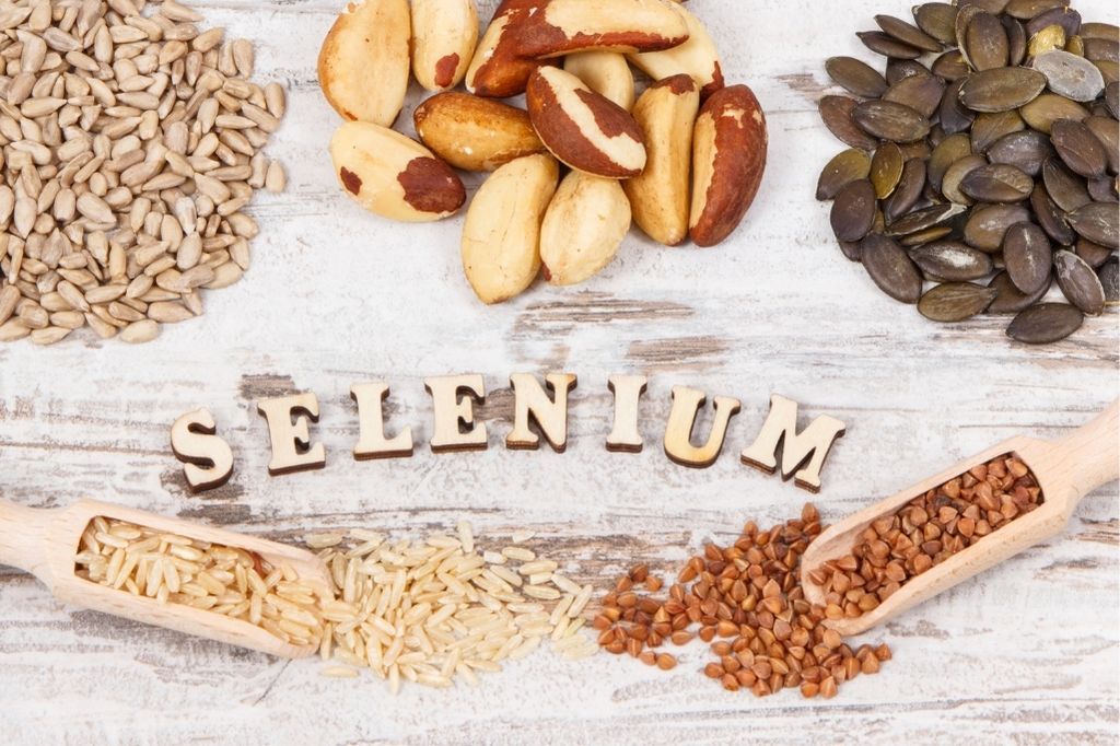
Selenium is an essential trace mineral needed in small quantities; without it we cannot live. Selenium is an antioxidant that helps protect cells from damage caused by oxidative stress. In addition to reducing oxidative stress, selenium supports proper immune function, and it seems to lower the risk of certain cancers. It is also necessary to produce enzymes. Brazil nuts are the best source of selenium. Other sources are fish, pork, beef, turkey and chicken, cottage cheese, eggs, brown rice, and sunflower seeds. Some processed foods are also enriched with synthetic selenium.
Zinc is a mineral found in cells throughout the body and is involved in cell division, cell growth, and wound healing. Zinc is necessary for immune system function, sperm production, and carbohydrate metabolism. It is also needed for our sense of taste and smell, and it plays an important role in insulin use. Zinc is essential for fetal development. It is also essential for growth and development of infants and children.
The Importance Of Magnesium
Lastly, manganese is a precursor to many enzymes, and it is needed for protein digestion and absorption as well as cholesterol metabolism. It is important for bone health, and, when taken with calcium, zinc and copper, it helps reduce spinal bone loss in older women. Manganese is necessary for blood sugar regulation, and it functions as an antioxidant. It is a key element for wound healing, and it does so by aiding in the production of proline, an amino acid necessary for collagen production. Manganese is found in a variety of foods including oysters, clams and mussels, nuts, legumes and unrefined grains, coffee, tea, and many spices. Our body stores up to 20 mg of manganese in the kidneys, liver, pancreas and bones.
What is the RDA level for each of the minerals? What is the upper limit for each of the vitamins? What are the signs or symptoms of deficiency and toxicity for each of the minerals? (300 words)
The recommended dietary amount for iodine is age dependent. A newborn requires 110 mcg of iodine daily. An infant between 7 to 12 months requires 130 mcg. Children between 1 and 8 years require 90 mcg, and children between 9 and 13 require 120 mcg. Teenagers between 14 and 18 require 150 mcg daily, which is the same for adult males and females. Pregnant women require at least 220 mcg daily, and lactating women require 290 mcg. Excess iodine intake causes fever, nausea, vomiting, weak pulse, stomach pain, and a burning sensation in the throat, mouth, and stomach, and coma. Chronic iodine toxicity is rare but can result in thyroid gland inflammation and even thyroid cancer. Iodine deficiency causes inadequate production of thyroid hormones which leads to hypothyroidism. Iodine deficiency can also cause the thyroid gland to enlarge, causing goiter. Iodine deficiency increases risk of miscarriage, stillbirth, and congenital abnormalities in babies. Tolerable upper limit intake level (UL) has also been established for iodine. This is the maximum amount of iodine that can be safely ingested without risking toxicity/overdose. The UL for iodine intake is as follows: children between 1 and 3 have a UL of 200mcg; for children between 9 and 13 UL is 600mcg; UL is 900mcg for teenagers between 14 and 18 years, and it is 1100mcg for adults.
Selenium Deficiency
Selenium deficiency is rare in the general population, but cases of selenium deficiency are often reported in patients who receive intravenous feeding therapy for extended periods of time. As a result, patients are likely to develop Keshan disease, a cardiomyopathy that presents with enlargement of the heart muscle resulting in congestive cardiac failure, cardiogenic shock and death. Another condition caused by selenium and iodine deficiency is Kashin-Beck disease. Kashin-Beck is prevalent mostly in China, Siberia, Korea and Tibet and is caused by mineral deficiencies associated with depleted soil.
It affects joints and bones to the point that limbs’ growth are stunted, joints are deformed, and the individual experiences loss of stature caused by necrosis of growth plates of the bones and of the cartilage in the joints. Cognitive function is also affected and mental retardation can be present. Excess selenium in the body results in selenosis, which causes fatigue, nausea, mild nerve damage, disrupted vision, nail problems, and hair loss. The RDA for selenium vary based on age, gender, illnesses, pregnancy, and they are as follows:
Category
Age
Units in mcg/day
Infants
Up to six months
15
7 – 12 months
20
Children
1 – 3 years
20
4 – 8 years
30
9 – 13 years
40
Adolescents and adults
Above 14 years
55
Pregnant women
–
60
Lactating women
–
70
Signs and Symptoms Of Zinc Deficiency
The signs and symptoms of zinc deficiency include poor appetite, stunted growth, alopecia, skin sores, wounds that do not heal easily, hypogonadism, poor olfactory and taste senses, and poor vision. Similarly, a high zinc intake (usually through supplements) causes vomiting, diarrhea, and abdominal cramps within three to ten hours. These symptoms generally stop once supplementation ends. Zinc toxicity has antagonistic effects on copper and iron. The RDA for zinc are established to prevent deficiency or toxicity and are as follows: infants between 0 and 6 months should take 2mg/day; infants and children 7 months to 3 years should take 3mg/day; RDA for children between 4 and 8 years is 5mg/day, and 8mg/day is recommended for children between 9 and 13 years. Males over 14 years should take 11mg/day, while females between 14 and 18 years should take 9mg/day. Females over 19 years of age require 8mg/day, 11mg/day during pregnancy and 12mg/day when lactating. Pregnant teenagers require 12mg/day, whereas those lactating require 13mg/day.
Manganese Deficiency Is Rare, But Its Absence Can Disrupt Healthful Living
Although rare, manganese deficiency is possible and manifests with retarded growth, infertility, skeletal abnormalities, low sugar tolerance, and altered fat and carbohydrate metabolism. Manganism or manganese poisoning results from chronic exposure to manganese and can manifest neurological disorders including tremors, facial muscle spasms or difficulty walking; these neurological symptoms are usually proceeded by hallucinations, reduced lung activities, and aggression. There is no RDA for manganese; however, the adequate intake (AI) is 1.8-2.3 mg/day, and the upper limit is 11mg/day for adults above 19 years.
What impact does the quality of the soil play in the role of the available minerals in our food supply? What types of chemicals in our environment impede mineral absorption?
Soil is an essential factor in healthy food and human and animal nutrition. While most of us are familiar with malnutrition that comes from lack or poor quality macronutrients, not everyone knows about another form of malnutrition called “hidden hunger”. Hidden hunger occurs when our diet is void of micronutrients, such as B vitamins, vitamin A, iodine, selenium, and zinc. Micronutrients are essential to health, being cofactors in metabolic functions, digestion, neurotransmitter production, eyesight, energy, and more. The Food and Agriculture Organization estimates that around 2 billion people worldwide suffer from hidden hunger; that is almost 1/3 of the world population. The quality of our soil is a key factor affecting micronutrient availability in food and nutrient quality.
Most the foods that we eat come from the soil: vegetables, fruits, grains and grains products. Hidden hunger is caused by lack of variety, consumption of crops that are not nutrient-rich, and soil depletion. Plants need 18 essential elements for proper growth and nutrient-density. Three of them (carbon, hydrogen and oxygen) are obtained through photosynthesis; the rest need to be present in the soil. Unsustainable soil management strips the soil of nutrients, causing the foods grown in it to have suboptimal nutrient profiles. We need to get away from mass farming procedures that encourage unsustainable soil management, and we need to embrace agricultural practices that are based on crop rotation and diversification which promote soil fertility; healthy soil leads to healthy food.
Healthy Soil Means A Healthy You
Another factor that plays an important role in soil health, and therefore the health of our crops, is exposure to chemicals, heavy metal pollution, and man-made fertilizers. Studies show that long-term use of man-made fertilizers causes a decline in soil quality and productivity. Particularly hazardous is long term exposure to heavy metals, insecticides and aromatic hydrocarbons. These substances have toxic effects on plants and humans; they prevent mineral absorption, disrupt vital enzymatic processes, change the microbiota, and compete with minerals for absorption. Cadmium, mercury, lead, arsenic and copper are the heavy metals most commonly found in the soil. They accumulate through disposal of high metal wastes, sewage sludge, wastewater irrigation, emissions from industrial areas, and spillage of petrochemicals.
How Cadmium Can Be Destructive For Healthy Living
Cadmium interferes with copper, iron and zinc. Mercury damages the nerves and interferes with cellular respiration. Lead ingestion can lead to anemia, kidney and brain damage and even death. Particularly problematic is exposure to lead by individuals whose diets are deficient in vitamin E, calcium, phosphorus, iron and zinc. Arsenic is a known carcinogen, which causes skin changes, nausea and vomiting, arrhythmia, and cramps. While there is no medication to combat arsenic poisoning, the use of vitamin E and selenium have shown to limit the symptoms and damage of arsenic poisoning.
Heavy metals do not only pose a risk to our health, but they are also harmful to soil regeneration and biodegradation of organic contaminants. Soil washing, immobilization, phytoremediation are all technologies available to clean up contaminated soils.
References
Stipanuk MH, Caudill MA, editors. Biochemical, physiological, and molecular aspects of human nutrition. 4th ed. St. Louis, Mo: Elsevier; 2019. 959 p.
Al-Fartusie, F. S., & Mohssan, S. N. (2017). Essential trace elements and their vital roles in human body. Indian J Adv Chem Sci, 5(3), 127-136.
Kavtarashvili, A. S., Stefanova, I. L., Svitkin, V. S., & Novotorov, E. N. (2017). Finctional egg production. II. The roles of selenium, zinc, and iodine. BIOLOGY AGRICULTURAL, 700.
Mishra, S., Bharagava, R. N., More, N., Yadav, A., Zainith, S., Mani, S., & Chowdhary, P. (2019). Heavy metal contamination: an alarming threat to environment and human health. In Environmental biotechnology: For sustainable future (pp. 103-125). Springer, Singapore.
Zimmermann, M. B., & Boelaert, K. (2015). Iodine deficiency and thyroid disorders. The Lancet Diabetes & Endocrinology, 3(4), 286-295.
What are the roles of lipoproteins and cholesterol in the body? Consider the interplay of insulin and cholesterol. How does a client’s insulin level impact his/her level of cholesterol? What is the current standard of care for someone who presents with elevated cholesterol? In what ways does this current standard of care affect insulin levels? Outline a nutritional protocol to help your client address his/her concerns of high cholesterol. Include labs might you request from your client’s primary care provider to assist you in designing this protocol. (600 words)
Lipids are hydrophobic: they are non-polar and insoluble in water. This means that they cannot dissolve in blood and rely on special particles for transport. These particles are called lipoproteins. Lipoproteins are a group of proteins synthesized in the small intestine and liver that transport hydrophobic lipids throughout the body. Lipoproteins are made up of lipids and proteins. The hydrophobic lipid portion of lipoproteins is placed in the core, while the hydrophilic protein portion is placed in the periphery of the particle. This particular structure is what allows lipoproteins to travel in the blood and transport lipids through the body.
Find Out What The Different Types of Lipoproteins and Their Names
There are different types of lipoproteins, and they are named according to the density of their content: chylomicron, chylomicron remnant, very low density lipoprotein (VLDL), intermediate density lipoprotein (IDL), low density lipoprotein (LDL), and high density lipoprotein (HDL). Chylomicrons are the least dense while HDL are the densest.
The lipids present in lipoproteins are triglycerides, phospholipids, free cholesterol, and cholesterol ester. Cholesterol is a high-molecular-weight alcohol, and it comes from two sources: exogenous (dietary cholesterol contained only in food from animals) and endogenous (manufactured by the liver). Cholesterol has several vital functions within the body. It gives our cells stability and stiffness. It is a precursor for the synthesis of steroid hormones, vitamin D, and bile, and it acts as an antioxidant. Cholesterol is needed for serotonin function, and low levels of cholesterol have been linked to aggressive behavior, violence, depression, and suicidal tendencies. Breast milk is rich in cholesterol, and infants and children need cholesterol-rich foods for proper development of the brain and nervous system. Cholesterol is also considered the “duct tape” of the body, used to repair damaged tissues.
Cholesterol and Heat Exposure
Cholesterol can become damaged upon exposure to heat and oxygen. Oxidized cholesterol is found in foods like fast foods, fried foods, margarines, baked goods, and foods that are deep fried in rancid vegetable oils.
Several studies reveal that prolonged exposure to insulin is linked to higher levels of lipid peroxidation markers in LDL. For this reason, we need to be aware that clients suffering from hyperinsulinemia will present higher levels of LDL compared to clients with normal blood sugar metabolism. The cholesterol guidelines from the American College of Cardiology and American Heart Association are as follows: patients with arterial plaques and otherwise healthy patients with LDL-C levels greater than or equal to 190 mg/dl are advised to drastically reduced intake of dietary cholesterol and are prescribed high-intensity statin therapy (or maximum tolerated statin therapy). Diabetic patients between the ages of 40 and 75 with LDL-C levels greater than or equal to 70 mg/ dl are prescribed moderate-intensity statin. It is disheartening to see such guidelines in place and to read that many expert physicians consider them not aggressive enough. Statins are dangerous medications linked to a host of side effects including memory loss and confusion, liver damage, muscle pain and damage. Statins also activate an immune response that prevents insulin from working correctly, causing an increase in blood sugar and, therefore, a higher incidence of diabetes. While as a nutritionist I cannot recommend against doctor’s orders, it is my duty to provide my clients with the latest research and information necessary to make informed decisions.
What Is High Cholesterol and How To Bring The Levels Down
When working with clients who are concerned about high cholesterol levels, some of the tests that I find helpful are the advanced lipid tests LDL particle number (LDL-P) and apolipoprotein B (apoB) as well as serum insulin test and c-reactive protein. These tests all measure biomarkers that can accurately predict risk of cardiovascular disease.
The nutritional protocol for such clients focuses on an anti-inflammatory diet that supplies high quality proteins, fats, and carbohydrates from low-glycemic vegetables and fruits. The diet removes added sugars, processed foods, fried foods, and vegetable oils. I also help them with stress management techniques and sleep hygiene. Supplements are an important part of nutritional therapy and, while there is no typical protocol, nutrients that are helpful in cases presenting high cholesterol are: chromium: 200-400 mcg with each meal; vitamin E: 200-600 IU d-alpha and d-gamma tocopherol; l-arginine: 700 mg two to three times a day with meals; magnesium orotate: starting with 400 mg and dosed to bowel tolerance; and curcumin: 15-60 mg three times a day. A formulation that I have used with success is Lipid-Sirt from Biotics Research.
References:
Naviglio D. Bad Cholesterol or “Bad” Science? Med chem [Internet]. 2016 [cited 2020 Oct 15];6(1). Available from: https://www.omicsonline.org/open-access/bad-cholesterol-or-bad-science-2161-0444-1000321.php?aid=66755
Shane Ellison — Life Saving Cholesterol Facts [Internet]. [cited 2020 Oct 15]. Available from: https://newswithviews.com/Ellison/shane13.htm
Kendrick M. The great cholesterol con: the truth about what really causes heart disease and how to avoid it. London: John Blake; 2008. 270 p.
Ross AC, editor. Modern nutrition in health and disease. 11th ed. Philadelphia: Wolters Kluwer Health/Lippincott Williams & Wilkins; 2014. 1616 p.
Shils ME, Shike M, editors. Modern nutrition in health and disease. 10th ed. Philadelphia: Lippincott Williams & Wilkins; 2006. 2069 p.
Smith LL. Another cholesterol hypothesis: cholesterol as antioxidant. Free Radic Biol Med. 1991;11(1):47–61.
Mei S, Gu H, Yang X, Guo H, Liu Z, Cao W. Prolonged Exposure to Insulin Induces Mitochondrion-Derived Oxidative Stress through Increasing Mitochondrial Cholesterol Content in Hepatocytes. Endocrinology. 2012 May 1;153(5):2120–9.
Colas R, Pruneta-Deloche V, Guichardant M, Luquain-Costaz C, Cugnet-Anceau C, Moret M, et al. Increased lipid peroxidation in LDL from type-2 diabetic patients. Lipids. 2010 Aug;45(8):723–31.
Grundy Scott M., Stone Neil J., Bailey Alison L., Beam Craig, Birtcher Kim K., Blumenthal Roger S., et al. 2018
Grundy SM, Stone NJ, Bailey AL, Beam C, Birtcher KK, Blumenthal RS, et al. 2018 AHA/ACC/AACVPR/AAPA/ABC/ACPM/ADA/AGS/APhA/ASPC/NLA/PCNA Guideline on the Management of Blood Cholesterol: A Report of the American College of Cardiology/American Heart Association Task Force on Clinical Practice Guidelines. Circulation [Internet]. 2019 Jun 18 [cited 2020 Oct 16];139(25). Available from: https://www.ahajournals.org/doi/10.1161/CIR.0000000000000625
This website collects cookies. Please read our Privacy Policy to review the updates about which cookies we use and what information we collect on our site. By continuing to use this site, you are agreeing to our updated privacy policy.